Wearing contact lenses gives patients the flexibility and freedom to live life to the fullest, without some of the difficulties presented by wearing glasses. Many people who choose contact lenses do so because they don’t like the way that glasses look or feel, or because wearing glasses compromises their ability to perform certain tasks or activities, such as sports or jobs that require the use of safety goggles.
There are lots of different contact lenses to choose from, with two of the most popular being daily disposables and toric lenses.
Disposable Lenses
As their name suggests, these daily contact lenses are disposable. This means that they can and should be discarded at the end of each day rather than re-worn. Disposable lenses do tend to be a little more expensive than some repeat-wear varieties, but the benefits usually outweigh the cost.
Some of the advantages of choosing daily disposable contact lenses include:
You don’t have to clean them, which saves patients a great deal of time and hassle. It also helps save money in terms of the ongoing cost of cleaning solution.
Disposable lenses are also great for people with eye allergies. This is because with ordinary lenses, there’s an opportunity for deposits and microorganisms to build up. With daily disposables, allergens have less chance to attach themselves to the lenses and cause irritation and other allergy symptoms.
You don’t need to schedule regular replacements either, which makes wearing contact lenses easier on your schedule.
Disposable contact lenses are particularly good for people who have busy lives and are likely to cut corners when it comes to caring for their eyes or contacts since there is no cleaning or maintenance required.
Daily disposable contact lenses are available in a wide range of prescriptions, including those for patients with nearsightedness and farsightedness. Your eye doctor will be able to advise you if you are a candidate for disposable contact lenses.

Neuro-Optometric Rehabilitation
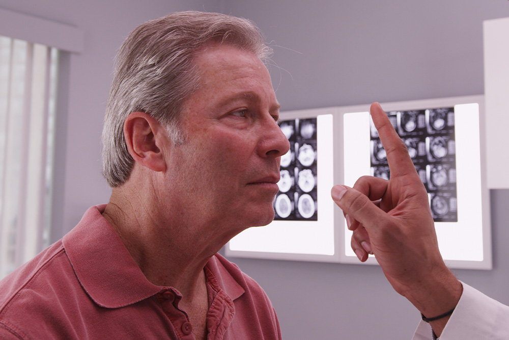
When most people think about vision problems, they picture issues with the eyes themselves. However, many visual difficulties actually begin in the brain. After a concussion, stroke, or other brain injury, the connection between the eyes and brain can be disrupted - causing symptoms that affect balance, reading, focus, and even daily comfort.
Brain Injuries and Vision
Your visual system is complex - about 70% of the brain is involved in vision processing. After a traumatic brain injury (TBI), concussion, or neurological event such as a stroke, the pathways that coordinate eye movement, focus, and perception can become impaired.
This can result in symptoms such as:
- Blurred or double vision
- Eye strain and headaches
- Difficulty reading or focusing
- Dizziness or balance problems
- Light sensitivity
- Problems with depth perception
These visual issues often persist long after other symptoms of a brain injury have improved, making specialized vision care essential for recovery.
What Is Neuro-Optometric Rehabilitation?
Neuro-optometric rehabilitation is a specialized form of vision therapy designed to retrain how the brain and eyes work together. Unlike traditional eye care that focuses on eyesight alone, this therapy targets the neurological processes behind vision. Our doctor evaluates how the eyes and brain communicate and creates a personalized treatment plan to rebuild these visual pathways. The goal is to restore comfort, coordination, and clarity - improving overall visual function and quality of life.

Dry eye is a common condition that occurs when the eyes do not produce enough tears or when the tears evaporate too quickly. There are several factors that can contribute to dry eye, including environmental factors, hormonal changes, and certain medications. Additionally, conditions such as meibomian gland dysfunction and blepharitis can also lead to dry eye.
The Role of Meibomian Gland Dysfunction and Blepharitis in Dry Eye
Meibomian gland dysfunction occurs when the meibomian glands, which are responsible for producing the oily layer of tears, become blocked or do not function properly. This can result in the tears evaporating too quickly and not providing enough lubrication for the eyes. Blepharitis is an inflammation of the eyelids that can cause redness, itching, and a gritty sensation in the eyes. Both meibomian gland dysfunction and blepharitis can contribute to dry eye by disrupting the natural tear film.
Recognizing the Symptoms of Dry Eye
Dry eye can cause a variety of symptoms, which can vary in severity from person to person. Some common symptoms of dry eye include:
Dryness: The most common symptom of dry eye is a feeling of dryness or grittiness in the eyes. This can make it uncomfortable to wear contact lenses or spend long periods of time looking at a screen.
Redness: Dry eye can cause the blood vessels in the eyes to become more prominent, leading to redness and irritation.
Itching: Some people with dry eye may experience itching or a burning sensation in their eyes.
Excessive tearing: Dry eye can also cause excessive tearing. This is the body's response to the irritation caused by the lack of lubrication in the eyes.
If you are experiencing any of these symptoms, it is important to consult with an optometrist for a proper diagnosis and treatment plan.

Dry eyes are one of the most common conditions that can affect our eyes and is estimated to affect millions of Americans. As you’ve probably guessed, dry eyes occur when tears fail to provide enough natural lubrication for the eyes to be comfortable and healthy. Exactly what causes dry eyes can vary significantly, from side effects from medications to prolonged computer use. What is clear is that while the condition isn’t sight-threatening, it can make day to day life much harder than it needs to be. Fortunately, there are treatments that can help, and arguably one of the most effective is Lipiflow.
What is Lipiflow?
Lipiflow is a new technological solution that addresses the underlying cause of your dry eyes, rather than simply treating the symptoms. It is most effective at helping patients whose dry eyes are caused by meibomian gland dysfunction – a condition characterized by problems with the way that the meibomian glands produce the oil that forms an essential part of our tear film. The meibomian glands can become less productive, or in some cases, even blocked by hardened oil deposits. This prevents the oil from reaching your tear film, making it less effective. Lipiflow targets the meibomian glands, warming them to break down oily blockages and massaging your eyes to make sure that the oil, and then the tear film, is evenly dispersed. This helps to combat the symptoms associated with dry eyes, which can include:
Eye fatigue
Dry, scratchy and uncomfortable eyes
Blurred vision
Sensitivity to light
Difficulty wearing contact lenses
Your eye doctor will be able to advise you if Lipiflow has the potential to be a suitable solution for your dry eyes.

Whether people like it or not, fine lines and wrinkles go hand in hand with the aging process. However, they typically appear sooner and look worse for those who spend time in the sun. Fortunately, TempSure Envi offers a solution that improves the skin’s appearance and health.
Instead of having invasive surgery, experts in the field of aesthetics can offer their patients something better. Not only is TempSure Envi non-invasive, but it’s also safe and effective. Simply put, it provides optimal improvement without any pain or discomfort.
Beyond Fine Lines and Wrinkles
Eliminating fine lines and wrinkles is just one of many benefits associated with TempSure Envi treatments. This same treatment works incredibly well to reduce the appearance of cellulite. Overall, it smooths skin, making it look more youthful.
However, even leading ophthalmologists and optometrists rely on TempSure Envi to treat patients with dry eye disease. Usually caused by Meibomian Gland Disease or MGD, the combination often makes a person look tired. In addition to dealing with uncomfortable symptoms, this causes bags to form beneath the eyes.
Because TempSure Envi is a gentle and safe treatment, it’s ideal for giving people with dry eye disease from MGD a fresher appearance.
How Does TempSure Envi Work?
This treatment uses a radiofrequency that gently and safely heats the skin for a specific amount of time. The body naturally reacts by producing new collagen. Because the new fibers are tighter and denser, they fill in voids in the form of lines, wrinkles, and cellulite. It also diminishes bagginess associated with dry eye disease from MGD.

A tonometer refers to the equipment that is used in tonometry – a test that measures the pressure inside your eyes, also known as intraocular pressure or IOP for short. Tonometry is rarely performed at your average comprehensive eye exam unless you are at high risk of or have been already diagnosed with glaucoma. Fortunately, tonometry can be used to detect changes in eye pressure before they cause any symptoms, enabling prompt action to be taken before your vision is affected.
About Glaucoma
Glaucoma is a common eye condition that occurs when the optic nerve, which connects the eye to the brain, becomes damaged. It’s normally caused by fluid building up in the front part of the eye, which causes the pressure inside the eyes to build. As the pressure increases, the optic nerve becomes increasingly damaged, and this prevents messages from being transmitted between your eyes and brain effectively. As a result, the patient’s vision becomes compromised. Without treatment, the level of vision loss will continue to increase. Unfortunately, any vision that has been lost as a result of glaucoma cannot be restored.
Most of the time, glaucoma develops very slowly which means that many people don’t realize that they are affected until some damage to their vision has already occurred. However, occasionally glaucoma can develop quickly, and symptoms do occur.
Glaucoma symptoms can include:
Red eyes
Intense headaches
Tenderness around the eyes
Eye pain
Seeing rings/halos around lights
Blurred vision
Nausea and vomiting
If you notice any of these symptoms, it’s important that you make an appointment with your eye doctor right away so that you can be assessed. You are likely to have a tonometry test as part of this assessment.

Urgent eye care encompasses prompt evaluation and treatment of sudden or severe eye-related issues, including foreign object removal, chemical exposure, corneal abrasions, sudden vision loss, eye trauma, acute glaucoma, chemical burns, and eye infections. Seeking immediate professional attention from an optometrist is vital to prevent further damage and preserve vision.
Common Eye Emergencies or Urgent Eye Care Appointments
Eye emergencies can manifest in various forms, and it is essential to be able to identify them quickly. Some common eye emergencies include:
Foreign Object in the Eye: Particles, debris, or small objects can become lodged in the eye, causing pain, redness, tearing, and potential damage to the eye's surface.
Corneal Abrasions or Scratches: Injuries to the cornea, such as abrasions or scratches, can cause severe eye pain, light sensitivity, and a feeling of something in the eye.
Sudden Loss of Vision: Any sudden and unexplained loss of vision requires immediate attention to determine the underlying cause and initiate appropriate treatment.
Eye Trauma or Blunt Force Injury: Injuries to the eye from impact, trauma, or accidents can lead to serious complications, including retinal detachment, hemorrhage, or intraocular foreign bodies.
Chemical Burns: Exposure to caustic substances or chemicals can cause serious damage to the eyes, resulting in pain, redness, and potential vision loss.
Eye Infections: Infections such as conjunctivitis (pink eye) can cause redness, discharge, and discomfort in the eyes.
Recognizing these symptoms and seeking urgent care can prevent further complications.
The Importance of Basic Red Eye Exams in Urgent Care
Red eye exams are a fundamental part of urgent eye care. They help identify the cause of redness and determine the appropriate treatment. Basic red eye exams involve a comprehensive evaluation of the eye, including examining the eyelids, conjunctiva, cornea, and iris. These exams aid in the diagnosis and treatment of various conditions, such as conjunctivitis, uveitis, dry eyes, and corneal abrasions.

In September 2025, the FDA approved Stellest lenses for children ages 6 to 12 to correct and manage myopia, a major milestone for myopia care. These lenses have been successfully used in countries across Europe, Canada, and Asia since 2020, showing promising results in slowing eye growth associated with myopia.
The Importance of Early Myopia Management
Myopia typically begins in early childhood, often between the ages of 6 and 10, and tends to worsen quickly during the school years as children spend more time reading and using digital devices. The sooner it begins, the higher the risk that it will progress to more severe levels. Every increase in myopia level significantly raises the risk of eye health complications later in life, such as retinal detachment or glaucoma.
By beginning myopia management as soon as possible, parents can help slow eye growth and protect long-term vision. With innovative options like Stellest lenses, families now have effective, science-backed solutions to take control of their child’s myopia.
What Are Stellest Lenses?
Stellest lenses are a groundbreaking innovation in myopia control, developed by Essilor. They look like regular eyeglass lenses but are designed with specialized optical technology to both correct vision and slow the progression of myopia.

If you find it difficult to tell colors apart, you may be color blind. Color blindness, or color deficiency, is estimated to affect around 8% of men and about 1% of women, but for those affected, it can significantly impact the quality of their day-to-day life. Contrary to popular belief, being color blind doesn’t mean that you can’t see any color at all. Instead, patients simply struggle to differentiate between certain colors. The vast majority of people who are color blind find it impossible to tell the difference between varying shades of red and green. You may hear this referred to as red-green color deficiency. However, this doesn’t only mean that they mix up red and green. They can also mix up colors that have some green or red light as part of their whole colors, for example purple and blue. This is because they are unable to see the red light that forms part of the color purple.
As you can probably imagine, this type of visual impairment can be a problem for things like traffic lights, taking medications and even looking at signs and directions. For example, someone who is color blind may find that the green on a traffic light may appear white or even blue.

When it comes to maintaining your eye health, regular check-ups and screenings play a crucial role. One such screening method that has revolutionized the field of optometry is retinal imaging testing. This non-invasive procedure allows optometrists to capture detailed images of the retina, providing valuable insights into the overall health of your eyes.
The Importance of Retinal Imaging Testing
By examining the retina, which is the thin layer of tissue at the back of your eye responsible for capturing light and transmitting visual signals to the brain, optometrists can gain valuable insights into your eye health. Retinal imaging testing allows for the early detection of various eye conditions even before noticeable symptoms occur. This early detection is crucial as it enables prompt treatment and intervention, potentially preventing irreversible vision loss.
Advanced Retinal Imaging Technology
Retinal imaging testing has been made possible by the advancements in technology, specifically Optical Coherence Tomography (OCT) and Optos imaging. OCT is a non-invasive imaging technique that uses light waves to capture high-resolution cross-sectional images of the retina. It provides detailed information about the layers of the retina, helping eye doctors identify and monitor various eye conditions.
Optos imaging utilizes ultra-widefield retinal technology to capture a panoramic image of the retina. This technology allows for a more comprehensive view of the retina, including the periphery. Dilating drops are not necessary with Optos, making the process more convenient for patients.


